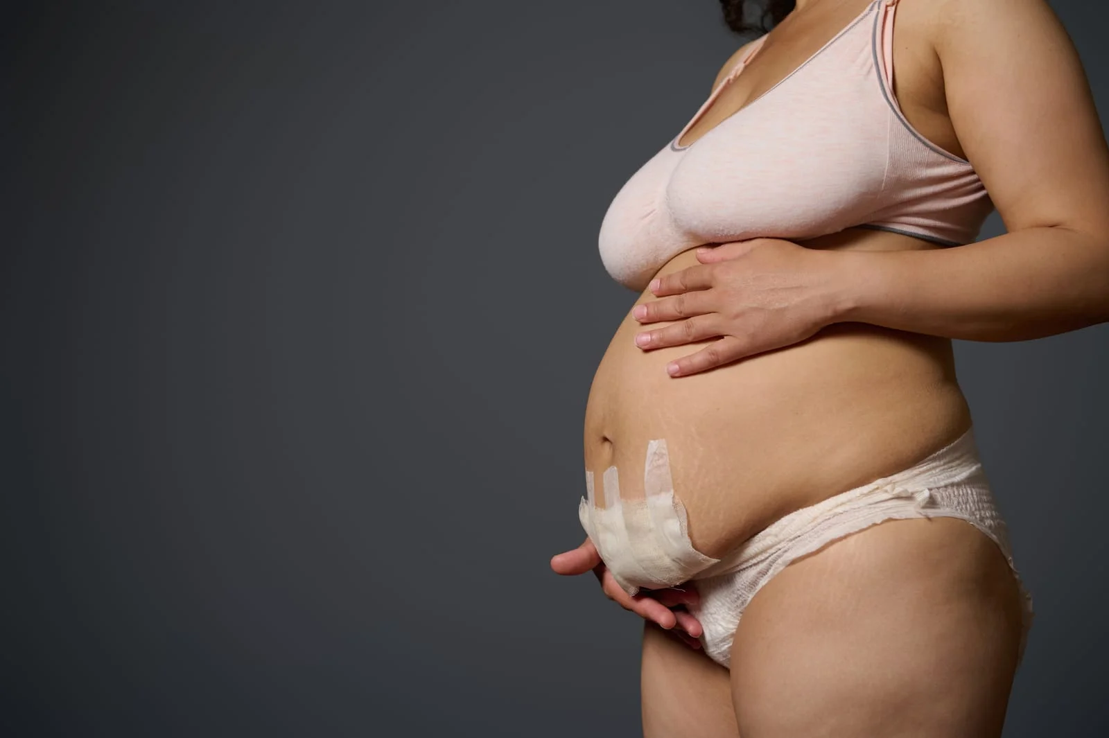
Cesarean sections currently account for 20 to 30% of all births, a figure that is steadily rising as the procedure becomes a common necessity for both emergency and planned deliveries. The Institute of Human Anatomy contributes to a deeper public understanding of this surgery by utilizing cadaveric dissections to demonstrate the complex layers of tissue and muscle that must be navigated to reach a fetus. Because no universal procedure exists, surgeons must adapt their approach based on the mother’s health, body habitus, and the urgency of the birth. In most planned cases, a low transverse incision—commonly known as the "bikini line cut"—is utilized, strategically placed two to five centimeters above the pubic symphysis to avoid the bladder and provide an aesthetically discrete result.
The surgical journey begins with an incision through the epidermis, dermis, and the hypodermis, a subcutaneous layer of fatty tissue that varies significantly in thickness between patients. To facilitate faster healing and reduce the risk of severing blood vessels, surgeons often transition to blunt dissection, using their hands or the blunt ends of instruments to reveal the deeper tissues rather than relying solely on a scalpel. Beneath the fat lies the rectus sheath, a white connective tissue fusion created by the sheet-like tendons, or aponeuroses, of the oblique and transversus abdominis muscles. Rather than cutting through the "six-pack" muscle (rectus abdominis), surgeons target the linea alba, a vertical midline of connective tissue. By making a vertical incision here, they can simply separate the muscle heads to expose the abdominal cavity without causing excessive muscle damage.
Related article - Uphorial Shopify

Inside the cavity, the surgeon encounters the peritoneum, a thin serous membrane that must be breached to reveal the uterus. During a full-term pregnancy, the uterus is a massive organ that has typically pushed the greater omentum (a fatty apron) and the small intestines toward the sides or behind it. The uterine incision is generally transverse and positioned near the infant's head to facilitate the delivery of the baby, the placenta, and the amniotic sac. Once the delivery is complete, the uterus is sutured closed; in some techniques, it may even be temporarily lifted out of the pelvic cavity to allow for more precise stitching before being returned to its position.
The final phase of the C-section focuses on restoring the body's structural integrity. While the closure of the peritoneum and subcutaneous fat is often at the surgeon's discretion, it is essential to suture the linea alba back together. This step is critical because it restores the strength of the abdominal wall and prevents the future formation of hernias. From the initial skin incision to the final stitch, the procedure illustrates the remarkable adaptability of the human body, particularly the uterus, which can grow to reach near the sternum to accommodate a developing child.
Undergoing a C-section is much like carefully unzipping a multi-layered winter jacket to reach a fragile package tucked inside the lining; by following the natural seams and zippers of the body rather than cutting through the fabric itself, the surgeon ensures the contents are safely removed while the jacket remains functional and ready to be repaired.


